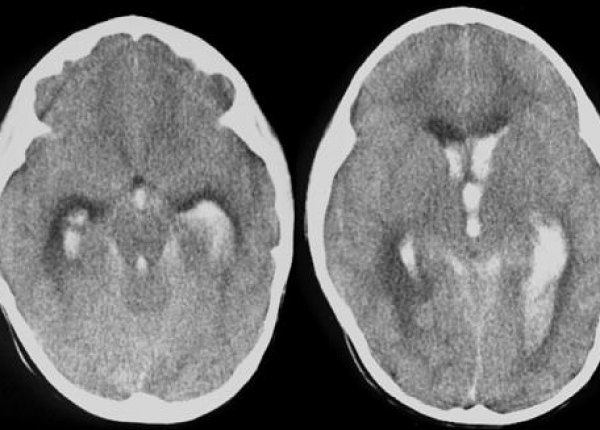
Neuroimaging of White Matter Injury, Intraventricular and Cerebellar Hemorrhage

White matter injury and hemorrhage are common findings in extremely preterm infants. Large hemorrhages and extensive cystic lesions are identified with cranial ultrasound. MRI, which is more sensitive, is especially useful in the identification of small intraventricular hemorrhage; cerebellar hemorrhage; punctate lesion in the white matter and cerebellum; and diffuse, noncystic white matter injury.
Imaging sequences such as diffusion-weighted, diffusion tensor, and susceptibility weighted imaging may improve recognition and prediction of outcome. These techniques improve understanding of the underlying pathophysiology of white matter injury and its effects on brain development and neurodevelopmental outcome.
Manon J.N.L. Benders, Karina J. Kersbergen, and Linda S. de Vries
تصویربرداری عصبی از آسیب نسج سفید، داخل بطن و خونریزی مخچه ای
آسیب نسج سفید و خونریزی، یافته های شایعی در نوزادان خیلی نارس می باشند. خونریزی های بزرگ و ضایعات کیستیک وسیع توسط سونوگرافی مغز تشخیص داده میشوند. MRI بخصوص در تشخیص خونریزی های داخل بطنی کوچک، خونریزی مخچه ای، ضایعات نقطه ای در نسج سفید و مخچه و آسیبهای منتشر غیر کیستیک نسج سفید حساستر می باشد.توالیهای
تصویری همچون diffusion-weighted, diffusion tensor ممکن است تشخیص و پیش بینی عاقبت بیمار را بهتر انجام دهد. این تکنیکها فهم پاتوفیزیولوژی زمینه ای اسیب نسج سفید و اثرات ان را روی تکامل مغزی و سرانجام تکامل عصبی بهبود می بخشد.
(اصل مقاله در مرکز تحقیقات سلامت نوزادان موجود است)

